Synthesis on a coarse-grained ribosome
The Continuous Synthesis Protocol (CSP) is cosmo’s codon-resolved, kinetic runner
for protein synthesis on an explicit coarse-grained ribosome. It times
every residue from its mRNA codon and splits each elongation cycle into three
kinetic sub-stages — reproducing O’Brien’s continuous_synthesis_v6.py protocol, but
on cosmo’s sequence-based IDP force field (HPS / mpipi) instead of a structure-based
Gō model. (Codon-resolved kinetics are what make the model physically meaningful; a
fixed per-residue step count is not.) For the analytic-tunnel variant — the same codon
kinetics with the explicit ribosome replaced by a cylindrical bore — see
Synthesis through an analytic tunnel (cylinder model).
CLI:
cosmo-csp -f csp.ini(orpython -m cosmo.csp -f csp.ini)Movie tool:
cosmo-csp-movie -o <out_root> [--ribosome ribo.pdb]Worked example:
tutorials/08_csp_cg_ribosome/— a tutorial-scaled run on α-synuclein grown on the real E. coli 4V9D CG ribosome (the analytic-tunnel variant istutorials/07_csp_cylinder/). A larger production configuration lives insandbox/validate/andsandbox/Ecoli/.Architecture: CSP is a thin outer loop. The per-length MD work — building the length-
Lmodel, seeding coordinates, restraints, running one stage — lives in the shared low-level enginecosmo.csp.core(run_length,RunParams); the rigid-ribosome scenery and tunnel wall live incosmo.csp.ribosome; the timing lives incosmo.csp.kinetics. CSP adds only the kinetics and the three-run_length-calls-per-residue loop.
Note
This is the cosmo port of topo.csp. It mirrors the sibling topo package’s module
layout, INI keys and CLI, but the nascent chain is an intrinsically disordered protein
under the HPS / mpipi force field — so there is no STRIDE, no native-contact map, no
domain.yaml, and no build-once-subset machinery. A length-L model is simply
cosmo.models.buildCoarseGrainModel on the first L residues of the sequence.
Quick start
All paths in the INI are relative to the working directory; run from the example folder. A GPU is recommended for production (the explicit-ribosome system has ~4,600 rigid beads).
cd tutorials/08_csp_cg_ribosome
cosmo-csp -f csp.ini              # 3-stage synthesis -> synth_out_csp/

# stitch the per-stage trajectories into one VMD movie
cosmo-csp-movie -o synth_out_csp --ribosome 4v9d_50S_PtR_5jte_AtR_model_cg_trunc.pdb
cd synth_out_csp && vmd -e movie.tcl        # movie.tcl loads its files by basename
cosmo-csp writes, per residue L and sub-stage s, a standalone trajectory under
<outdir>/L_<L>/ (one folder per residue, per-stage traj_s<s>.dcd), optional stall/ and ejection/ phases, and a
per-residue dwell-time log <outdir>/dwell_times.dat.
Theory
1. What is being modeled: protein synthesis
In a living cell a protein is synthesized vectorially, N-terminus first, by the ribosome, one amino acid at a time, while the growing (“nascent”) chain threads out through the ribosomal exit tunnel (~80 Å long, ~10–20 Å wide) and begins to fold or collapse co-translationally — as it emerges. The kinetics of synthesis matter: how long the ribosome dwells on each codon sets how much time each segment of the chain has to sample conformations before the next residue is added. Rare codons (decoded slowly) act as “translational pauses”. CSP reproduces this by growing a coarse-grained protein bead-by-bead out of a coarse-grained ribosome, timing each residue from its mRNA codon.
Because cosmo’s chain is an IDP (no native fold), CSP here studies the co-translational behaviour of disordered chains — extrusion, tunnel confinement, compaction — rather than folding toward a native structure.
The real elongation cycle (one amino acid added)
Bacterial translation elongation repeats a three-step biochemical cycle per codon:
Aminoacyl-tRNA selection / decoding. A ternary complex delivers an aa-tRNA to the ribosomal A site; correct codon–anticodon pairing triggers accommodation. This is the codon-dependent, highly variable, usually rate-limiting step (cognate-tRNA abundance / codon-usage bias).
Peptidyl transfer. The peptidyl-transferase center (PTC) transfers the nascent peptide from the P-site tRNA onto the A-site aminoacyl-tRNA. The chain is now one residue longer and attached to the A-site tRNA. Fast (~0.3 ms).
Translocation. EF-G ratchets the ribosome forward by one codon: the tRNAs move A→P, the A site is freed. ~few ms.
CSP partitions the per-codon dwell time into these three pieces and reproduces them as three MD sub-stages per residue.
2. The simulation model (one elongation step)
Nascent protein — a sequence-based IDP chain. One bead per residue at the Cα position. Interactions are set by the sequence, not a structure: the short-range Ashbaugh–Hatch (HPS) or Wang–Frenkel (mpipi) pair potential plus Debye–Hückel electrostatics. There are no native contacts — the chain does not fold toward a target structure. Bonds are flexible harmonic.
Ribosome — rigid scenery. The truncated CG 50S + tRNAs (~4,600 mass-0 beads) is fixed in space, but its excluded-volume and electrostatic interactions with the nascent chain are on. The tunnel axis is aligned with +x (the chain exits +x).
The PTC anchors. Two ribosome beads are singled out as fixed reference points: the P-anchor (P-site tRNA residue-76
Rbead) and the A-anchor (A-site tRNA residue-76Rbead) — where the peptidyl-tRNA (P) and incoming aminoacyl-tRNA (A) hold the chain’s C-terminus.C-terminus restraint — position restraint (default) or O’Brien tRNA tether. The current C-terminal bead is held at the A-/P-site either by a harmonic position restraint to the target point (
U = k·|r − r₀|²,k = restraint_k = 83680 kJ/mol/nm²= 200 kcal/mol/Ų; default) or, withtrna_tether = yes, by the full O’Brien tRNA tether — a bond + two orienting angles + an improper to the A76 tRNA beads (plus a backbone orienting angleprev–N–R— thehps_ssbistable backbone potential, applied across the peptide–tRNA junction for all models), which also fixes the chain’s orientation. Switching the A-/P-site hold A→P is how translocation is reproduced — the position restraint moves its target point; the tether re-attaches to the P-site beads in stage 3. (Thekis a per-particle parameter so this force coexists with the tunnel wall, whose global constant is alsok.)Tunnel wall — a one-sided plane. Because the 50S is truncated to a shell around the exit tunnel, there are no ribosome beads below the PTC. A one-sided half-harmonic wall
U = k·min(x − x₀, 0)²(fixed stiffness 8368 kJ/mol/nm² = 20 kcal/mol/Ų) supplies the missing floor: the chain can only extrude forward (+x) and cannot slip below the synthesis point into the truncated region. The planex₀is auto-derived from the ribosome structure (the lower of the two C-terminus hold planes,min(A-target.x, P-target.x)), so it can never go stale when you switch structures.Thermostat. Langevin dynamics at
ref_t = 300 K, frictiontau_t, timestepdt.
Length-L model (no build-once-subset). cosmo’s forces are all sequence-local or
pairwise-by-type, so the length-L model is exactly
buildCoarseGrainModel on residues 1..L of the sequence — bonds, Yukawa, the
short-range HPS/mpipi term (and, for hps_ss, the local angle/torsion). Going L → L+1
just adds residue L+1’s terms. No STRIDE, no contact map, no matrix injection.
3. Ribosome ↔ nascent excluded volume: the O’Brien 12-10-6
The rigid ribosome interacts with the nascent chain through excluded volume + electrostatics only — no attractive/native contacts. The excluded volume is O’Brien’s 12-10-6 form (inherited verbatim from topo):
U = ε·[13(R/r)¹² − 18(R/r)¹⁰ + 4(R/r)⁶] ,   R = Rmin/2ᵢ + Rmin/2ⱼ  (sum rule)
ε = 0.000132 kcal/mol ,   cutoff 2.0 nm / switch 1.8 nm ,   {nascent}×{ribosome} only
The per-bead Rmin/2 (O’Brien’s structure-based CG collision radii) are
model-independent steric radii: they live in the standalone OBRIEN_RMIN_2_NM
(per-amino-acid) and OBRIEN_RNA_RMIN_2_BEADS (rRNA P/R/BR) tables in
cosmo.parameters.model_parameters, decoupled from any force field. So the ribosome
wall is identical for every nascent model — CSP runs on hps_kr, hps_urry or
mpipi alike (hps_kr is merely the default); the selected model only sets the
nascent IDP↔IDP interaction (Ashbaugh–Hatch or Wang–Frenkel). Electrostatics fold into the existing
Yukawa force, extended over the ribosome charges (rRNA phosphate −1e, charged
residues) on {nascent}×{nascent} + {nascent}×{ribosome} (no intra-ribosome
electrostatics; the rigid ribosome’s own interactions are constant and never computed).
4. The three stages: biology ↔ simulation
Each amino acid is added through one elongation cycle, split into three kinetic
sub-steps; CSP runs one MD segment per sub-step. For nascent length L each sub-stage
is a short simulation writing its own traj_s<s>.dcd in the shared L_<L>/ folder; stage 3’s final
structure seeds the next residue’s stage 1.
stage |
real process |
what the simulation does |
C-terminus restrained to |
mean dwell |
|---|---|---|---|---|
1 |
Peptidyl transfer |
new bead |
A-target |
|
2 |
Translocation (onset) |
continue from stage 1, still held at the A-target; run MD (minimize skipped) |
A-target |
|
3 |
Translocation completes + wait |
switch the restraint A→P, then run MD |
P-target |
remainder = (next codon total) − stage 1 − stage 2 |
Note
Mechanics vs. timing. The restraint switch (an instantaneous A→P geometric move)
happens at the start of stage 3, while the duration charged to translocation is
stage 2. With the default position restraint only the C-terminus point is held;
explicit A/P tRNA bonded geometry (bond + angles + improper to the A-/P-site tRNA beads,
switched A→P across the stages) is modelled when trna_tether = yes. The timing
(three codon-resolved dwell times per residue) is faithful to O’Brien; the per-stage
mechanics are a reduced model.
4a. C-terminus restraint: position restraint vs. tRNA tether
The C-terminus must be held at the PTC and translocated A→P each residue. cosmo offers two
mechanisms, chosen by the trna_tether key (see Synthesis control options); both drive the
same A→P schedule across the three stages, but the tether additionally controls the
chain’s orientation.
Position restraint (trna_tether = no, default). A single harmonic spring pins the
C-terminal bead to the A/P target point (the equilibrium-optimized A- or P-site location),
U = k·|r − r₀|². Stages 1–2 target the A-point, stage 3 targets the P-point; the A→P
switch is just changing r₀. This is the validated default — simplest, and it needs no
particular tRNA beads.
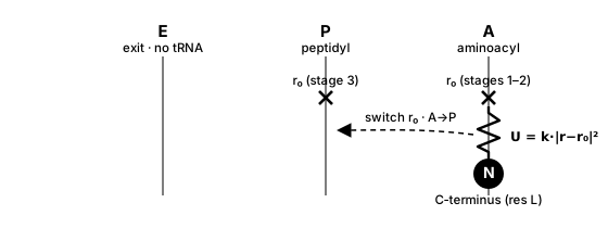
Position restraint (trna_tether = no). A single harmonic spring pins the C-terminal
bead to the A- or P-site target point; translocation is just moving the target r₀ from
the A-point (stages 1–2) to the P-point (stage 3) — right→left in the E–P–A frame.
O’Brien tRNA tether (trna_tether = yes). Reproduces the covalent attachment of the
nascent C-terminus to the tRNA with the full bonded geometry — not a point restraint but a
set of bonded terms to the tRNA’s 3′-terminal nucleotide, residue 76 (A76).
Which residue? The truncated ribosome keeps only the acceptor-stem 3′ end of each tRNA
(a handful of residues: AtR 73–76, PtR 72–76), each nucleotide represented by a few CG
beads — P (phosphate), R (ribose), BR1/BR2 (base). The tether attaches to residue
76 only — the 3′-terminal adenosine A76, which is exactly the CCA-3′ site where the amino
acid esterifies to the tRNA. The other tRNA residues are rigid excluded-volume scenery,
not part of the tether. It uses three beads of A76:
R— the ribose of A76: the C-terminus bonds to it.P— the phosphate of A76: used in the orienting angleN–R–P.BR2— the second base bead of A76 (present only because A76 is a purine): used inN–R–BR2and the improper.
For the current C-terminus N (the newest residue) it adds:
term |
force |
connectivity (beads of A76) |
A-site ( |
P-site ( |
|---|---|---|---|---|
bond |
harmonic |
|
0.427 nm |
0.476 nm |
angle |
harmonic |
|
106° |
117° |
angle |
harmonic |
|
127° |
130° |
improper |
periodic ( |
|
128° |
−161° |
backbone |
double-Gaussian angle |
|
the |
(bond/angle stiffness = 200 kcal/mol/Ų; angle/improper = 25 kcal/mol/rad².) The two orienting angles + the improper fix the residue’s bearing in the A76 frame — this is what the plain point restraint cannot do.
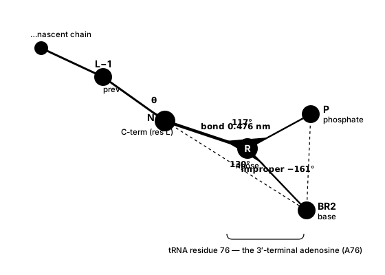
tRNA tether geometry (trna_tether = yes). The C-terminus (N) bonds to the
ribose (R) of A76; two orienting angles (N–R–P, N–R–BR2) and the improper
(N–R–P–BR2) fix its bearing in the A76 frame, and a backbone angle (prev–N–R) keeps the
terminal segment physically oriented (the hps_ss double-Gaussian backbone potential, added
for all models). Values shown are the P-site (PtR) set;
A-site (AtR) values are in the table above. Only A76 is tethered — the rest of the tRNA
is rigid scenery.
Per-stage tethering (tRNA-tether mode). Each stage rebuilds the length-L system and
re-attaches the tether to the appropriate site — there is no sliding spring; the A→P
“switch” is realized by the tether referencing the P-site beads (PtR) instead of the
A-site beads (AtR):
stage |
C-terminus (residue |
previous residue ( |
|---|---|---|
1 |
A-site ( |
P-site ( |
2 |
A-site ( |
— free — |
3 |
P-site ( |
— free — |
Stage 1 double-tethers both ends of the freshly-formed peptide bond (the new residue at A, the previous one at P), so the new bond starts at its equilibrium PTC geometry the instant the residue appears.
Stage 2 keeps the new residue at A; the previous residue is released (held only by the nascent-chain backbone + the tunnel wall).
Stage 3 re-tethers the new residue A→P — starting from stage 2’s coordinates, the minimize + MD carries it from the A-site to the P-site: this is the translocation.
Cold start (the first residue
L0, no previous residue): tethered to the P-site in all three stages.
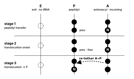
The A→P switch across the three stages. Each stage rebuilds the system and re-attaches
the tether (no sliding spring). Stage 1 double-tethers both ends of the new peptide bond
(new residue N at A, previous at P); stage 2 keeps N at A (previous released); stage 3
re-tethers N to the P-site — the minimize + MD carry it A→P (right→left), this is the
translocation. Sites are drawn E–P–A per convention; CSP models only the P- and A-site
tRNAs (no E-site tRNA).
Note
The tRNA tether requires a well-formed A/P tRNA in the ribosome PDB (segids AtR/PtR,
resid 76, beads R/P/BR2; the acceptor must be a purine, which carries BR2). A site
missing its P or BR2 bead simply skips that angle/improper. The A/P target points
(used by both modes, and for the tunnel-wall plane) come from the always-on PTC-geometry
optimization.
5. From codon to MD steps (the kinetics)
The timing core is cosmo.csp.kinetics (pure Python, no OpenMM), identical to topo’s.
For every residue it answers: how many integration steps does each sub-stage run?
(a) Per-codon mean translation time. The mRNA is split into codons; a lookup table
maps each codon to its mean in-vivo translation time in seconds — the codon’s
intrinsic mean first-passage time (mFPT), τ(codon).
Note
The codon-time table is an organism + temperature property (not of the protein), so a
per-codon run needs an explicit codon_times table path — there is no bundled default.
The shared library under assets/csp/codon_dwell_times/<organism>/ holds tables per
organism, including the Fluitt E. coli table at 310 K (Fluitt, Pienaar & Viljoen,
Comput. Biol. Chem. 2007; 61 sense + 3 stop codons, codon-usage-weighted mean ≈ 0.061 s
≈ 16.5 aa/s) at assets/csp/codon_dwell_times/ecoli/ecoli_codon_dwell_times_310K.txt. Both
mrna (the protein-specific sequence) and codon_times (the table) are mandatory for
per-codon timing; see Codon dwell-time tables (per-codon timing).
(b) First-passage-time sampling. Each stage is gated by a single rate-limiting event,
so its waiting time is exponentially distributed. Each sub-stage’s dwell is drawn as
t = −mean·ln(U), U ∼ Uniform(0,1). A fixed random_seed makes the schedule
reproducible.
Important
time_stage_1 / time_stage_2 (and the stage-3 remainder) are means, not the
per-residue dwell. The actual time on each residue is a fresh exponential draw with that
mean, so individual residues get shorter/longer dwells and only their average equals the
configured value.
(c) The three-stage split for nascent length L (1-indexed). Draw three independent
U₁, U₂, U₃:
t1(L) = −time_stage_1                                  · ln(U₁)   # peptidyl transfer
t2(L) = −time_stage_2                                  · ln(U₂)   # translocation
t3(L) = −(τ(next codon) − time_stage_1 − time_stage_2) · ln(U₃)   # wait to decode next codon
Stage 3 uses the next codon’s mean (having just added residue L, the ribosome now
waits for L+1’s tRNA — which is why the codon list must extend to L_max + 1). If a
fast next codon makes the remainder ≤ 0 it is floored to 1e-9 s.
(d) In-vivo seconds → in-silico steps. A time-compression factor maps seconds to steps:
t_sim (ns) = t_s · 1e9 / scale_factor
n_steps    = t_sim (ns) / dt(ns) ,   dt(ns) = dt_ps · 1e-3
A larger scale_factor ⇒ fewer steps per residue ⇒ a faster run, while preserving the
relative timing of fast vs. slow codons. Step counts may additionally be clamped to
[min_steps_per_stage, max_steps_per_stage] for tractability — a clamp on MD steps
only; the sampled dwell times in seconds are recorded untouched in dwell_times.dat.
6. PTC geometry (always optimized)
The A-site seed / stage-1-2 restraint target and the P-site / stage-3 target are placed
by optimal_ptc_targets exactly one peptide bond apart (cosmo’s CG bond length —
0.380 nm for hps_kr, not topo’s 0.381 nm) and clear of the ribosome excluded
volume, by minimizing the soft O’Brien tRNA-bond/angle/improper restraints + the 12-10-6
wall over a deterministic multistart. Each new residue is delivered with its peptide bond
at equilibrium, which drops the stage-1 potential energy by ~50× versus seeding at the raw
AtR/PtR-76 R anchor beads (which sit ~0.9 nm apart, badly stretching the bond). By default the
C-terminus is held by a position restraint to these points; with trna_tether = yes
it is instead bonded to the A-/P-site tRNA beads (the full O’Brien tether). The PTC-target
optimization itself is always on; there is no knob for it.
Note
cosmo defaults to flexible harmonic bonds (constraints = None) because backbone
flexibility is physically meaningful for disordered chains, but it also supports rigid
bonds (constraints = AllBonds) — every CA/P bond becomes a distance constraint pinned
at its equilibrium length, removing the fast bond-stretch mode so a larger dt can be
used. Constraints act only on the bonds; the non-bonded potentials are untouched.
So AllBonds does not by itself prevent the stiff-EV blow-up: the non-native
excluded volume is still stiff — the new residue is seeded at the fixed A-site target,
and a recently-added residue that has not cleared that region can land in the repulsive
core of the ribosome↔nascent 12-10-6 EV or the nascent Ashbaugh-Hatch potential (both
present in cosmo and topo). That diverges at the configured dt (PotE → ~1e13 kJ/mol),
which is why cosmo keeps topo’s per-stage dt-halving stability guard: it re-runs the
stage at dt/2 with 2× steps (identical dwell) until it is stable. The always-on PTC
optimization reduces how often this fires (equilibrium seeding lowers the stage-1 energy
~50×), but it is a quality improvement, not a full substitute for the guard.
7. After the last residue: ejection
Once the final residue is added, up to two post-synthesis phases run in order. First,
an optional stall phase (stall_steps) keeps the finished chain at the PTC with the
C-terminus restraint / tRNA tether still on — a ribosome-stalling hold. Then the
ejection phase (ejection_steps) releases the restraint while the rigid ribosome
and tunnel wall remain, so the chain diffuses out along +x. Each phase is skipped when its
step count is 0.
The ejection is extendable, but extension is opt-in via restart (default no):
ejection_steps is the cumulative free-run length. If the chain has not fully left the
ribosome, set restart = yes, raise ejection_steps, and re-run on the existing outdir
(with resume = auto) — the ejection continues from its checkpoint (positions, velocities
and step count) and appends only the additional steps, exactly like resuming a simulation
(if the checkpoint already reached the target it reports “already met” and does nothing).
With the default restart = no, a completed ejection is instead skipped on resume
(like the stall phase) — raising ejection_steps does not re-run or extend it; use
restart = yes (or resume = no to redo the whole synthesis). Inspect
ejection/traj_final.pdb yourself and top it up until the
chain is clear; the nascent-only final is then a ready ribosome-free input for a
post-translation cosmo-mdrun.
Configuration
CSP reads a single INI control file with one [OPTIONS] section
(cosmo.csp.protocol.read_csp_config). Every control key — for both cosmo-csp and
cosmo-cylinder — is documented in one place: Synthesis control options (grouped into
shared, coarse-grained-ribosome-only, and cylinder-only keys, with types and
defaults). Units are OpenMM defaults (nm, ps, kJ/mol, K, kJ/mol/nm²) and dwell times are
in seconds.
A few CSP-specific behaviors worth calling out (the physics behind the keys):
pdb_filemay be all-atom or a Cα-only CG structure — only the Cα positions and residue names are read (cosmo has no STRIDE / native-contact step), so there is nodomain_def/stride_output_file/nascent_ev_radiikey (those are topo’s Gō-model inputs).PTC geometry is always optimized (A/P targets one peptide bond apart, EV-clear), so each new residue is delivered with its bond at equilibrium — no knob.
The ribosome is always rigid scenery (supplying the
ribosomePDB is the signal; norigid_ribosomekey) and output is always nascent-only.trna_tetherdefaults off (the switchable A↔P position restraint); settrna_tether = yesfor the full O’Brien tRNA tether, which switches A→P by re-attaching to the P-site beads in stage 3.
Outputs
<outdir>/
├── L_<L>/                  # ONE folder per residue L (consolidated layout)
│   ├── traj.psf            # nascent topology (shared across the 3 stages; f(L) only)
│   ├── native_1_<L>.pdb    # length-L native structure (shared)
│   ├── traj_s1.dcd         # (nascent-only) trajectory, stage 1  (s2/s3 likewise)
│   ├── traj_s1.log         # energies, stage 1
│   ├── traj_runinfo.log    # folded run-info: one [run:...]/[result:...] per stage
│   └── traj_final.pdb      # stage-3 final — seeds L+1 and is the resume-reload target
├── stall/                  # post-synthesis stall phase, held at PTC (if stall_steps > 0)
├── ejection/               # post-synthesis ejection phase (if ejection_steps > 0)
├── dwell_times.dat         # per-residue dwell-time log / schedule (#PTC header)
└── progress.log            # append-only DONE/RUNNING resume status
Each residue’s three sub-stages share one L_<L>/ directory: traj.psf and
native_1_<L>.pdb depend only on L and are written once; trajectories stay split per
stage (traj_s{1,2,3}.dcd); only stage 3 writes traj_final.pdb. There is no per-stage
.chk — per-residue resume reloads traj_final.pdb. Re-invoking cosmo-csp on an
interrupted <outdir> continues from the last completed residue (resume = auto by
default; --fresh to override); see cosmo.csp.resume.
dwell_times.dat records, per residue, the codon, the three sampled dwell times in
seconds (t1/t2/t3), their nanosecond equivalents, and the integer MD step counts —
the physical schedule, independent of any step clamp.
Console progress log
cosmo-csp prints one compact line per residue plus one per sub-stage, each reporting the
wall-clock time and the total system potential energy of the last integrated step:
L=  5  uniform  dwell      0.05 s  steps   40/  40/  40
  L=  5  stage 1 peptidyl-transfer       40 steps    0.15 s  PE=  +6.7544e+01 kJ/mol
  L=  5  stage 2 translocation           40 steps    0.11 s  PE=  +5.9xxxe+01 kJ/mol
  L=  5  stage 3 tRNA-binding            40 steps    0.14 s  PE=  +5.6842e+01 kJ/mol
Set COSMO_CSP_VERBOSE=1 to restore the full per-stage banners (build block,
minimization, run metadata). MDAnalysis’ cosmetic warnings are silenced for the run.
Movie. Each stage writes a standalone trajectory; cosmo-csp-movie stitches them — in
synthesis order, padding every frame to the final length and overlaying the static
ribosome — into one VMD-playable movie (auto-detects the 3-stage vs flat layout):
cosmo-csp-movie -o <outdir> --ribosome 4v9d_50S_PtR_5jte_AtR_model_cg_trunc.pdb
cd <outdir> && vmd -e movie.tcl        # movie.tcl loads its files by basename
For the cylinder-tunnel equivalent and more detail, see Visualizing the synthesis process.
Python API
from cosmo.csp.protocol import run_continuous_synthesis, read_csp_config

# (a) drive it from an INI, exactly like the CLI:
cfg = read_csp_config("csp.ini")
run_continuous_synthesis(
    cfg.pdb_file, cfg.ribosome,
    L0=cfg.L0, L_max=cfg.L_max, out_root=cfg.outdir,
    mrna=cfg.mrna, codon_time_table_path=cfg.codon_time_table_path,
    params=cfg.params,
)

# (b) or construct parameters directly (the ribosome PDB is always rigid scenery;
#     the tunnel-wall plane is auto-derived from it):
from cosmo.csp.core import RunParams
params = RunParams(model="hps_urry",   # any IDP model works; hps_kr is the default
                   scale_factor=4331293.0, random_seed=1, ejection_steps=50000)
run_continuous_synthesis("asyn.pdb", "4v9d_50S_PtR_5jte_AtR_model_cg_trunc.pdb",
                         L0=5, L_max=10, mrna="mrna.txt", params=params)
See the API reference for the autodocumented modules.
See also
Synthesis through an analytic tunnel (cylinder model) — the analytic-tunnel variant (
cosmo-cylinder): the same codon kinetics with a nascent-only system and a single MD segment per residue.Synthesis control options — the concise
csp.inicontrol-options reference.cosmo.csp package — the API reference (
cosmo.csp.core,cosmo.csp.ribosome,cosmo.csp.kinetics).