Synthesis in coarse-grained ribosome model¶
The Continuous Synthesis Protocol (CSP) is the codon-resolved, kinetic runner for protein synthesis on an explicit coarse-grained ribosome. It times every residue from its mRNA codon and splits each elongation cycle into three kinetic sub-stages — the published continuous-synthesis protocol, in topo style. (Codon-resolved kinetics are what make the model physically meaningful; a fixed per-residue step count is not.) For the analytic-tunnel variant — the same codon kinetics with the explicit ribosome replaced by a cylindrical bore — see Synthesis in cylinder ribosome model.
CLI:
topo-csp -f csp.ini(orpython -m topo.csp -f csp.ini)Movie tool:
topo-csp-movie -o <out_root> [--ribosome ribo.pdb]Worked example: Tutorial B.2 (
tutorials/B2_ribosome_synthesis/) — a smoke run (csp_debug.ini, L = 1 → 8) and a full-length validation (csp_val.ini, L = 1 → 306) on 4c5c, plus a second protein (P0CX28).Architecture: CSP is a thin outer loop. The per-length MD work — building the length-
Lmodel, seeding coordinates, restraints, running one stage under the stability guard, build-once-subset contacts — lives in the shared low-level enginetopo.csp.core(run_length,RunParams); the rigid-ribosome scenery and tunnel wall live intopo.csp.ribosome; the timing lives intopo.csp.kinetics. CSP adds only the kinetics and the three-run_length-calls-per-residue loop. The force field and ribosome representation are described in the Theory section below.
Quick start¶
All paths in the INI are relative to the working directory; run from the tutorial folder. A GPU is recommended (the explicit-ribosome system has ~4,600 ribosome beads).
# Run the CSP on 4c5c (Tutorial B.2)
cd tutorials/B2_ribosome_synthesis/4c5c
topo-csp -f csp_debug.ini       # smoke run -> synth_out_debug/  (or csp_val.ini -> synth_out/ for full length)

# Stitch the per-stage trajectories into one VMD movie
topo-csp-movie -o synth_out_debug --ribosome ribosome_trunc.pdb
topo-csp writes one folder per residue L under <outdir>/L_<L>/ (a shared topology
plus a per-stage trajectory traj_s<s>.dcd), optional stall/ and ejection/ phases, and a
per-residue dwell-time log <outdir>/dwell_times.dat.
Theory¶
1. What is being modeled: protein synthesis¶
In a living cell a protein is synthesized vectorially, N-terminus first, by the ribosome, one amino acid at a time, while the growing (“nascent”) chain threads out through the ribosomal exit tunnel (~80 Å long, ~10–20 Å wide) and begins to fold as it emerges. The kinetics of synthesis matter: how long the ribosome dwells on each codon sets how much time each segment of the chain has to sample conformations before the next residue is added. Rare codons (decoded slowly) act as “translational pauses” that can change folding outcomes.
CSP reproduces this: it grows a coarse-grained protein bead-by-bead out of a coarse-grained ribosome, timing each residue from its mRNA codon, so the nascent chain folds under realistic, codon-resolved kinetics.
The real elongation cycle (one amino acid added)¶
Bacterial translation elongation repeats a three-step biochemical cycle per codon:
Aminoacyl-tRNA selection / decoding. A ternary complex (EF-Tu·GTP·aminoacyl-tRNA) delivers an aa-tRNA to the ribosomal A site. Correct codon–anticodon pairing triggers GTP hydrolysis and accommodation. This is the codon-dependent, highly variable, usually rate-limiting step — its duration depends on cognate-tRNA abundance (codon-usage bias).
Peptidyl transfer (peptide-bond formation). The peptidyl-transferase center (PTC) transfers the nascent peptide from the P-site tRNA onto the A-site aminoacyl-tRNA. The chain is now one residue longer and attached to the A-site tRNA. Fast (~0.3 ms).
Translocation. EF-G·GTP ratchets the ribosome forward by one codon: the tRNAs move A→P and P→E, the deacylated tRNA leaves via the E site, and the A site is freed for the next aa-tRNA. ~few ms.
CSP partitions the per-codon dwell time into exactly these three pieces and reproduces them as three MD sub-stages per residue.
2. The simulation model (one elongation step)¶
Nascent protein — a structure-based (Gō-like) coarse-grained chain. One bead per residue at the Cα position. Native contacts (residue pairs close in the folded crystal structure) get attractive wells; everything else is repulsive — so the native structure is the energy minimum and the growing chain folds toward its native fold. Bonds default to rigid
AllBondsconstraints (stable because each residue is seeded at its equilibrium bond length — see §6); setconstraints = Nonefor flexible harmonic bonds instead.Ribosome — rigid scenery. The truncated CG 50S + tRNAs (~4,600 mass-0 beads) is fixed in space, but its excluded-volume and electrostatic interactions with the nascent chain are on, so the chain feels the tunnel walls and ribosome surface. The tunnel axis is aligned with +x (the chain exits toward +x).
The PTC anchors. Two ribosome beads are singled out as fixed reference points: the P-anchor (P-site tRNA residue-76 “R” bead) and the A-anchor (A-site tRNA residue-76 “R” bead). They stand in for where the peptidyl-tRNA (P) and incoming aminoacyl-tRNA (A) hold the chain’s C-terminus.
C-terminus restraint — two modes. The current C-terminal bead is held at the A- or P-site, and switching that hold A→P is how translocation is reproduced. There are two mechanisms, selected by
trna_tether(see §3a for the full details):Position restraint (
trna_tether = no, default): a single harmonic spring to the A/P target point,U = k·|r − r₀|²,k = restraint_k = 83680 kJ/mol/nm²(= 200 kcal/mol/Ų).O’Brien tRNA tether (
trna_tether = yes): the full covalent attachment to the tRNA — a bond + two orienting angles + an improper + a backbone angle to the actual A-/P-site tRNA beads, which also fixes the chain’s orientation in the tRNA frame (aiming it down the tunnel), not just its position.
Tunnel wall — a one-sided plane on the nascent beads. The rigid ribosome we supply is truncated to a shell around the exit tunnel (§8), so the model has no density on the −x (synthesis-interface) side of the PTC. In the real ribosome that side is not empty — it is occupied by the small subunit, the mRNA and the A-/P-site tRNAs, so the nascent chain can never move there — but truncation leaves an unreal void that the flexible nascent chain could drift backward (−x) into and tangle. A single one-sided half-harmonic restraint on every nascent bead,
U = k·min(x − x₀, 0)²(k = 8368 kJ/mol/nm²= 20 kcal/mol/Ų, a fixed model constant), supplies the missing barrier: it pushes any bead atx < x₀back tox ≥ x₀, so the chain can only extrude forward (+x, toward the cytosolic exit) and cannot slip below the synthesis point into the truncated region. The planex₀is auto-derived from the ribosome structure, not a config knob: it sits at the lower (smaller-x) C-terminus hold plane,x₀ = min(A-target.x, P-target.x)— the P-site, where the C-terminus is tethered — so the held C-terminus sits on the plane and the chain grows away from it. It is recomputed for whatever ribosome PDB you supply, so it never goes stale; for Tutorial B.2’sribosome_trunc.pdbit is x₀ ≈ 1.05 nm.Thermostat. Langevin dynamics at
ref_t = 310 K, frictiontau_t = 0.05 /ps, timestepdt = 0.015 ps.Optional flexible exit-tunnel loop. The ribosome is rigid by default, but
ribo_free_maskcan free a portion of the exit-tunnel protein (E. coli L24 / eukaryotic L26) as a mobile structure-based Go loop — the β-hairpin lining the tunnel can then breathe and contact the nascent chain. See the flexible-loop notes.
Build-once-subset contacts. The contact map is computed once on the full native
structure (R_full, eps_full, N×N). For nascent length L, the model uses the
top-left L×L block — STRIDE and the heavy-atom contact analysis are never re-run per
length. So residues 1..L carry exactly the native contacts they will have in the final
fold, and the chain can form native structure as soon as the relevant residues exist.
3. The three stages: biology ↔ simulation¶
Each amino acid is added through one ribosome elongation cycle, split into three
kinetic sub-steps; CSP runs one MD segment per sub-step. For nascent length L, each
sub-stage is a short simulation writing its own traj_s<s>.dcd in the shared L_<L>/
folder; stage 3’s final structure seeds the next residue’s stage 1.
stage |
real biological process |
what the simulation does |
C-terminus restrained to |
mean dwell time |
|---|---|---|---|---|
1 |
Peptidyl transfer — peptide bond forms; chain now sits on the A-site tRNA |
new bead |
A-target |
|
2 |
Translocation (onset) — EF-G begins ratcheting forward |
continue from stage 1, still held at the A-anchor; run MD |
A-anchor |
|
3 |
Translocation completes + wait for next aa-tRNA |
switch the restraint A→P (this geometric move is the translocation), then run MD while the chain relaxes/folds |
P-anchor |
remainder = (next codon’s total) − stage 1 − stage 2 |
Note
Mechanics vs. timing. The restraint switch (an A→P re-hold applied at the start of
stage 3) is decoupled from the durations: the time charged to translocation is stage
2 and the decoding wait is stage 3. The peptide bond is present in the bonded model
from stage 1 rather than toggled on mid-stage. Explicit A/P tRNA bonded geometry (bond +
angles + improper to the tRNA beads) is modelled when trna_tether = yes (§3a); with
the default position restraint only the C-terminus point is held. The timing (three
codon-resolved dwell times per residue) is faithful to the reference protocol; the
per-stage mechanics are a reduced model.
3a. C-terminus restraint: position restraint vs. tRNA tether¶
The C-terminus must be held at the PTC and translocated A→P each residue. topo offers two
mechanisms, chosen by the trna_tether key (see Synthesis control options); both drive the
same A→P schedule across the three stages, but the tether additionally controls the
chain’s orientation.
Position restraint (trna_tether = no, default). A single harmonic spring pins the
C-terminal bead to the A/P target point (the equilibrium-optimized A- or P-site location),
U = k·|r − r₀|². Stages 1–2 target the A-point, stage 3 targets the P-point; the A→P
switch is just changing r₀. This is the validated default — simplest, and it needs no
particular tRNA beads.
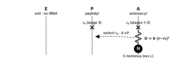
Position restraint (trna_tether = no). A single harmonic spring pins the C-terminal
bead to the A- or P-site target point; translocation is just moving the target r₀ from
the A-point (stages 1–2) to the P-point (stage 3) — right→left in the E–P–A frame.¶
O’Brien tRNA tether (trna_tether = yes). Reproduces the covalent attachment of the
nascent C-terminus to the tRNA with the full bonded geometry — not a point restraint but a
set of bonded terms to the tRNA’s 3′-terminal nucleotide, residue 76 (A76).
Which residue? The truncated ribosome keeps only the acceptor-stem 3′ end of each tRNA
(a handful of residues: AtR 73–76, PtR 72–76), each nucleotide represented by a few CG
beads — P (phosphate), R (ribose), BR1/BR2 (base). The tether attaches to residue
76 only — the 3′-terminal adenosine A76, which is exactly the CCA-3′ site where the amino
acid esterifies to the tRNA. The other tRNA residues are rigid excluded-volume scenery,
not part of the tether. It uses three beads of A76:
R— the ribose of A76: the C-terminus bonds to it.P— the phosphate of A76: used in the orienting angleN–R–P.BR2— the second base bead of A76 (present only because A76 is a purine): used inN–R–BR2and the improper.
For the current C-terminus N (the newest residue) it adds:
term |
force |
connectivity (beads of A76) |
A-site ( |
P-site ( |
|---|---|---|---|---|
bond |
harmonic |
|
0.427 nm |
0.476 nm |
angle |
harmonic |
|
106° |
117° |
angle |
harmonic |
|
127° |
130° |
improper |
periodic ( |
|
128° |
−161° |
backbone |
double-Gaussian angle |
|
the backbone-angle potential (bistable, θ ≈ 92°/130°) applied across the peptide–tRNA junction |
(bond/angle stiffness = 200 kcal/mol/Ų; angle/improper = 25 kcal/mol/rad².) The two orienting angles + the improper fix the residue’s bearing in the A76 frame — this is what the plain point restraint cannot do.
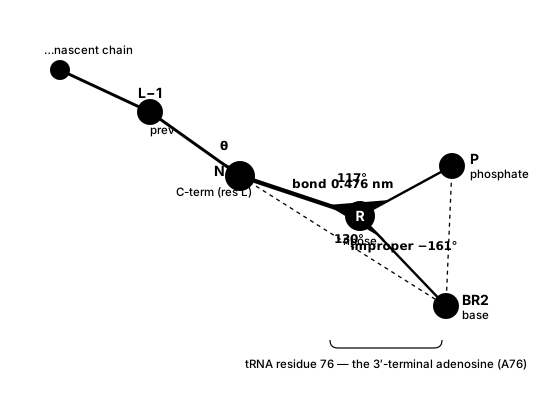
tRNA tether geometry (trna_tether = yes). The C-terminus (N) bonds to the
ribose (R) of A76; two orienting angles (N–R–P, N–R–BR2) and the improper
(N–R–P–BR2) fix its bearing in the A76 frame, and a backbone angle (prev–N–R) keeps the
terminal segment physically oriented. Values shown are the P-site (PtR) set; A-site (AtR) values are
in the table above. Only A76 is tethered — the rest of the tRNA is rigid scenery.¶
Per-stage tethering (tRNA-tether mode). Each stage rebuilds the length-L system and
re-attaches the tether to the appropriate site — there is no sliding spring; the A→P
“switch” is realized by the tether referencing the P-site beads (PtR) instead of the
A-site beads (AtR):
stage |
C-terminus (residue |
previous residue ( |
|---|---|---|
1 |
A-site ( |
P-site ( |
2 |
A-site ( |
— free — |
3 |
P-site ( |
— free — |
Stage 1 double-tethers both ends of the freshly-formed peptide bond (the new residue at A, the previous one at P), so the new bond starts at its equilibrium PTC geometry the instant the residue appears.
Stage 2 keeps the new residue at A; the previous residue is released (held only by the nascent-chain backbone + the tunnel wall).
Stage 3 re-tethers the new residue A→P — starting from stage 2’s coordinates, the minimize + MD carries it from the A-site to the P-site: this is the translocation.
Cold start (the first residue
L0, no previous residue): tethered to the P-site in all three stages.
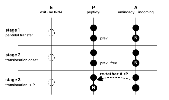
The A→P switch across the three stages. Each stage rebuilds the system and re-attaches
the tether (no sliding spring). Stage 1 double-tethers both ends of the new peptide bond
(new residue N at A, previous at P); stage 2 keeps N at A (previous released); stage 3
re-tethers N to the P-site — the minimize + MD carry it A→P (right→left), this is the
translocation. Sites are drawn E–P–A per convention; CSP models only the P- and A-site
tRNAs (no E-site tRNA).¶
Note
The tRNA tether requires a well-formed A/P tRNA in the ribosome PDB (segids AtR/PtR,
resid 76, beads R/P/BR2; the acceptor must be a purine, which carries BR2). A site
missing its P or BR2 bead simply skips that angle/improper. The A/P target points
(used by both modes, and for the tunnel-wall plane) come from the always-on PTC-geometry
optimization.
4. From codon to MD steps (the kinetics)¶
The timing core is topo.csp.kinetics (pure Python, no OpenMM). For every residue it
answers: how many integration steps does each sub-stage run?
(a) Per-codon mean translation time. The mRNA is split into codons; a lookup table
maps each codon to its mean in-vivo translation time in seconds — the codon’s
intrinsic mean first-passage time (mFPT). Call it τ(codon).
Note
What the codon-time table is. τ(codon) is the mean total per-codon translation
time — the full elongation-cycle mean first-passage time — and is dominated by, but
not equal to, the codon-dependent aminoacyl-tRNA decoding (selection) step (cognate-tRNA
availability / codon-usage bias). It is not only the decoding time: in CSP, τ is the
whole codon dwell, and stage 3 (τ − time_stage_1 − time_stage_2) is the remainder left
after the codon-independent peptidyl transfer and translocation — effectively the
decoding/tRNA wait.
The table is an organism + temperature property (not of the protein). A per-codon run
therefore needs an explicit codon_times table path – there is no bundled default. The
shared library under assets/csp/codon_dwell_times/ holds tables per organism, including
the Fluitt E. coli table at 310 K (Fluitt, Pienaar & Viljoen, Comput. Biol. Chem.
2007; 61 sense + 3 stop codons, mean ≈ 0.068 s ≈ 15 aa/s) at
assets/csp/codon_dwell_times/ecoli/ecoli_codon_dwell_times_310K.txt. Both mrna (the
protein-specific sequence) and codon_times (the table) are mandatory for per-codon timing.
Fastest / slowest / median (synonymous-codon) mRNA¶
Instead of supplying an mRNA file, you can have TOPO build one that keeps the protein
sequence fixed but reassigns each residue’s codon — a controlled walk along the
synonymous-mutation (codon-optimization) axis. Because the protein, its native structure,
and its contacts are identical across these mRNAs and only the elongation timing changes,
any difference in the co-translational folding they produce is attributable to codon
kinetics alone. Set mrna to a keyword instead of a filename:
fastest— the shortest-τsynonymous codon at every residue → the fastest translation the protein’s codons allow (minimises the mean total dwell time).slowest— the longest-τsynonymous codon at every residue → the slowest translation (maximises the mean total dwell); the slow codons act as translational pauses that give each emerging segment more time to fold.median— the middle-τsynonymous codon at every residue → a neutral reference sitting between the two extremes. When an amino acid has an even number of synonymous codons there is no single middle, so TOPO takes the faster (shorter-τ) of the two central codons; the pick is deterministic.
Here τ is the codon’s mean per-codon dwell time (introduced above), read from the
codon_times table — whose third column also supplies the codon→amino-acid map. The
runner reads the amino-acid sequence from pdb_file, picks one codon per residue as above,
appends the matching stop codon, and writes the raw-nucleotide mRNA next to the PDB as
mrna_fastest.txt / mrna_slowest.txt / mrna_median.txt; that file then feeds the normal
per-codon path. Because the pick is made per amino acid, the mode needs a codon-time
table — a single uniform codon_times number carries no synonymous choices and is
rejected. To pre-generate the file standalone (e.g. to inspect or version it):
topo-make-mrna --pdb protein.pdb \
    --codon-times assets/csp/codon_dwell_times/ecoli/ecoli_codon_dwell_times_310K.txt \
    --mode fastest
(b) First-passage-time sampling. Real elongation is stochastic: each stage is gated by a single rate-limiting molecular event, so its waiting time is exponentially distributed. Each sub-stage’s actual dwell time is therefore drawn at random as
t = −mean · ln(U) , U ∼ Uniform(0, 1)
(the inverse-transform draw of an exponential; the code uses the equivalent
random.expovariate(1/mean)). A fixed random_seed makes the whole schedule reproducible.
Important
time_stage_1 and time_stage_2 (and the stage-3 remainder) are means, not the
per-residue dwell. The values you set in csp.ini are the averages each stage
converges to over the chain; the actual time spent on each residue is a fresh random
draw from an exponential with that mean — so individual residues get shorter or longer
dwells (the exponential has a long tail: many short waits, occasional long ones), and only
their average equals the configured value. In the notation below, Exp(m) means “an
exponential whose mean is m” (the argument is the mean, not a rate). For example,
with time_stage_1 = 0.00034 s, draws of t1 scatter around 0.00034 s (≈0.00006 s when
U = 0.84, ≈0.00046 s when U = 0.26) and average to 0.00034 s.
(c) The three-stage split for nascent length L (1-indexed). Draw three independent
U₁, U₂, U₃ ∼ Uniform(0, 1) and compute the actual per-residue dwells:
t1(L) = −time_stage_1                                  · ln(U₁)   # peptidyl transfer
t2(L) = −time_stage_2                                  · ln(U₂)   # translocation
t3(L) = −(τ(next codon) − time_stage_1 − time_stage_2) · ln(U₃)   # wait to decode next codon
Each line is one sample from an exponential whose mean is the bracketed quantity —
i.e. t1(L) = −time_stage_1·ln(U₁) is the same statement as “t1 ∼ Exp(mean = time_stage_1)”. The bracketed quantities (time_stage_1, time_stage_2, and the stage-3
remainder) are the means; the t(L) values are the per-residue draws that scatter
around them. Example: residue L with U₁ = 0.84 gets
t1(L) = −0.000340·ln(0.84) ≈ 0.000057 s (this residue), while another residue with a
different U₁ gets a different t1 — and they average to time_stage_1 = 0.00034 s.
So the per-cycle clock = fixed-mean peptide bond + fixed-mean translocation + a
variable-mean decoding wait. (Indexing: stage 3 uses the next codon’s mean — having just
incorporated residue L, the ribosome now waits for residue L+1’s tRNA. This is why
build_codon_time_lists needs the codon list to extend to L_max + 1.)
(d) In-vivo seconds → in-silico steps. The coarse-grained model evolves far faster than real translation, so a time-compression factor maps seconds to MD steps:
t_sim (ns)  =  t_s · 1e9 / scale_factor
n_steps     =  t_sim (ns) / dt(ns) ,   dt(ns) = dt_ps · 1e-3
A larger scale_factor ⇒ fewer steps per residue ⇒ a faster run, while preserving the
relative timing of fast vs. slow codons (the physics that matters for folding during
synthesis). Step counts may additionally be clamped to
[min_steps_per_stage, max_steps_per_stage] for tractability — a clamp on MD steps
only; the sampled dwell times in seconds are recorded untouched in dwell_times.dat.
(e) Worked example (real 4c5c mRNA, Tutorial B.2). The first residues of
4c5c_mrna.txt and their τ from the default E. coli 310 K table:
residue |
codon |
τ (s) |
|---|---|---|
1 |
AUG |
0.133321 |
2 |
ACU |
0.027566 |
3 |
GAU |
0.038593 |
5 |
AUU |
0.048617 |
6 |
GCU |
0.019547 |
Take residue L = 1 (the ribosome has just made residue 1, codon AUG). Stage 3 looks
ahead to residue 2’s codon, ACU (τ = 0.027566 s), with time_stage_1 = 0.00034 s
and time_stage_2 = 0.004201 s:
mean(t3) = τ(ACU) − time_stage_1 − time_stage_2
         = 0.027566 − 0.00034 − 0.004201
         = 0.023025 s          # mean of the exponential; sampled t3 = −mean·ln(U)
Then → MD steps at Tutorial B.2’s scale_factor = 216564650, dt = 0.015 ps:
t_sim = 0.023025 s × 1e9 / 216564650 = 0.10632 ns
steps = 0.10632 ns / (0.015e-3 ns)   ≈ 7088 steps   (for the mean; then clamped to [400, 10000])
Stage 3 is where per-codon variability enters: stages 1 and 2 are fixed means, but
stage 3’s mean swings with the next codon’s τ (e.g. L = 5 → next codon GCU →
0.019547 − 0.00034 − 0.004201 = 0.015006 s). Edge case: if a very fast next codon makes
τ(next) − t1 − t2 ≤ 0, the mean is floored to 1e-9 s.
5. After the last residue: ejection¶
Once the final residue is added, the protein is complete. The simulation then runs up to
two post-synthesis phases in order. First, an optional stall phase (stall_steps)
keeps the finished chain at the PTC with the C-terminus restraint / tRNA tether still
on — a ribosome-stalling hold before release. Then the ejection phase
(ejection_steps) releases the restraint (the tether is cut), while the rigid ribosome
and one-sided tunnel wall remain. Biologically this is termination — release factors
free the finished protein. With the tether gone, the chain diffuses out of the tunnel
along +x (the one-sided wall biases motion forward) and clears the ribosome. Each phase
is skipped when its step count is 0. For a longer, dedicated egress demonstration, raise
ejection_steps in the Tutorial B.2 csp_val.ini.
The ejection is extendable, but extension is opt-in via restart (default no):
ejection_steps is the cumulative free-run length. If the chain has not fully left the
ribosome, set restart = yes, raise ejection_steps, and re-run on the existing outdir
(with resume = auto) — the ejection continues from its checkpoint (positions, velocities
and step count) and appends only the additional steps, exactly like resuming a simulation
(if the checkpoint already reached the target it reports “already met” and does nothing).
With the default restart = no, a completed ejection is instead skipped on resume
(like the stall phase) — raising ejection_steps does not re-run or extend it; use
restart = yes (or resume = no to redo the whole synthesis). Inspect
ejection/traj_final.pdb yourself and top it up until the
chain is clear; the nascent-only final is then a ready ribosome-free input for a
post-translation topo-mdrun.
6. Numerical integration, equilibrium seeding, and the stability guard¶
The default is rigid AllBonds at dt = 0.015 ps, and it is stable by construction.
This works because the always-on PTC-geometry optimization never pre-stretches a bond.
optimal_ptc_targets places the A- and P-site target points exactly one equilibrium
peptide bond apart — 0.381 nm, which is precisely the AllBonds constraint length —
and each new residue is seeded at the A-target, one equilibrium bond from the previous
C-terminus (which rests at the P-target). So the always-present peptide bond starts at
its rest length, and a rigid AllBonds build seeds and minimizes cleanly at 15 fs.
(Removing the fast bond-stretch mode is exactly how the reference protocol stays stable —
topo gets the same benefit by default.)
Note
This is a change from an earlier design in which the new residue was seeded far (~1 nm)
from its bond partner and flexible bonds were required to absorb the stretch. That is no
longer the case: the equilibrium-PTC seeding means the bond is never stretched, so rigid
AllBonds is the stable default. Flexible bonds (constraints = None) remain an
option — the equilibrium seeding keeps either treatment stable.
The stability guard is a safety net, not the primary mechanism. It mainly matters if
you switch to flexible bonds (constraints = None), where a newly-formed stiff native
(Gō) contact can drive a 15 fs step past stability and the dynamics diverge (potential
energy → ~10¹³ kJ/mol), corrupting that stage’s frames. The guard
(topo.csp.core.run_length) runs each stage in chunks while tracking the maximum
|PotE|; if a stage diverges (max |PotE| > 10⁹ kJ/mol) it is transparently re-run with
the timestep halved and the step count doubled — and because the dwell time is
n_steps · dt, halving dt and doubling n_steps leaves the dwell time exactly
unchanged (up to 6 halvings). Watch for [stability] … lines in the log. With the
default AllBonds seeding the guard essentially never fires: Tutorial B.2’s full-length run
(csp_val.ini, L = 1 → 306, AllBonds) completes all 919 stages with zero blow-ups.
Configuration¶
CSP reads a single INI control file — a flat key = value list, parsed by
topo.csp.protocol.read_csp_config. Every control key — for both topo-csp and
topo-cylinder — is documented in one place: Synthesis control options (grouped into
shared, coarse-grained-ribosome-only, and cylinder-only keys, with types and
defaults). Units are OpenMM defaults (nm, ps, kJ/mol, K, kJ/mol/nm²) and dwell times are
in seconds.
A few CSP-specific behaviors worth calling out here (the physics behind the keys):
PTC geometry is always optimized. Each new residue is seeded at the optimal A-site target — one equilibrium peptide bond (0.381 nm) from the previous C-terminus, clear of the ribosome excluded volume (
optimal_ptc_targets) — and those A-/P-site points are the restraint targets and the tunnel-wall plane. Because the peptide bond starts at equilibrium, rigidconstraints = AllBonds(the default) seeds and minimizes cleanly.The ribosome is always rigid scenery — supplying the
ribosomePDB is the signal (norigid_ribosomekey), andtrna_tetherdefaults off (CSP needs the switchable A↔P position restraint).
Note
tRNA presence / naming. The P-/A-anchors and the optimal_ptc_targets solve read the
ribosome’s tRNA beads under fixed names (segids PtR/AtR, resid 76, beads
R/P/BR2; the acceptor must be a purine A, which carries the BR2 bead). A ribosome
PDB with no tRNA, or with differently-named tRNA segments, currently fails with a
generic “expected exactly one bead” error. Handling missing/renamed tRNA is a tracked TODO
(review/TODO.md).
Outputs¶
<outdir>/
├── L_<L>/                  # ONE folder per residue L (consolidated layout)
│   ├── traj.psf            # nascent topology (shared across the 3 stages; f(L) only)
│   ├── native_1_<L>.pdb    # length-L native structure (shared; f(L) only)
│   ├── traj_s1.dcd         # (nascent-only) trajectory, stage 1  (s2/s3 likewise)
│   ├── traj_s1.log         # energies, stage 1; column 3 = potential energy (kJ/mol)
│   ├── traj_runinfo.log    # folded run-info: one [run:...]/[result:...] per stage
│   └── traj_final.pdb      # stage-3 final — seeds L+1 and is the resume-reload target
├── stall/                  # post-synthesis stall phase, held at PTC (if stall_steps > 0)
├── ejection/               # post-synthesis ejection phase (if ejection_steps > 0)
├── dwell_times.dat         # per-residue dwell-time log / schedule (see below)
└── progress.log            # append-only DONE/RUNNING resume status (see below)
Each residue’s three kinetic sub-stages share one L_<L>/ directory: traj.psf and
native_1_<L>.pdb depend only on L (the A/P differences are forces, not atoms) and are
written once; the trajectories stay split per stage (traj_s{1,2,3}.dcd) to preserve the
kinetic-dwell boundaries; and only stage 3 writes traj_final.pdb. There is no per-stage
.chk — per-residue resume reloads traj_final.pdb, never a checkpoint.
dwell_times.dat records, per residue, the codon, the three sampled dwell times in
seconds (t1/t2/t3), their nanosecond equivalents, and the integer MD step counts —
the physical schedule, independent of any step clamp. This is the file to compare against a
reference run for quantitative validation (Tutorial B.2). It is drawn once before the run
and doubles as the immutable plan for resume (its #PTC header pins the restraint
geometry); see Resuming long synthesis runs.
Resuming an interrupted run. A production synthesis is hours to days of wall time.
Re-invoking topo-csp on an interrupted outdir continues from the last completed
residue rather than restarting — the schedule and PTC geometry are re-read from
dwell_times.dat and the seed is reloaded from the last residue’s traj_final.pdb, tracked
by progress.log. Resume is on by default (resume = auto). See Resuming long synthesis runs
for the full mechanism, guarantees, and an HPC requeue pattern.
Console progress log¶
topo-csp prints one compact, column-aligned line per residue followed by one line per
sub-stage, so a long synthesis stays readable. Each stage line reports the wall-clock time
and the total system potential energy of the last integrated step (nascent chain +
rigid ribosome + all cross-interactions), a quick per-stage health signal:
L=  1    AUG  dwell   0.02757 s  steps  400/1136/2000
  L=  1  stage 1 peptidyl-transfer     400 steps    1.19 s  PE=  +4.9674e+01 kJ/mol
  L=  1  stage 2 translocation        1136 steps    0.42 s  PE=  +4.8717e+01 kJ/mol
  L=  1  stage 3 tRNA-binding         2000 steps    0.59 s  PE=  +4.8365e+01 kJ/mol
The residue line’s steps field and the per-stage steps are the configured step
counts; a stage that trips the stability guard silently reruns at a halved timestep with
double the steps (the [stability] ... lines above), and always prints its concise summary
line afterwards. The post-synthesis ejection phase prints the same summary line.
Set TOPO_CSP_VERBOSE=1 to restore the full per-stage banners (build block, seeded-
structure minimization, run-metadata path, elapsed time) — useful when debugging a single
length:
TOPO_CSP_VERBOSE=1 topo-csp -f csp.ini
MDAnalysis emits cosmetic UserWarnings (missing CRYST1 unit cell, absent
formalcharges) each time topo slices and writes a CA-only PDB — once per stage. topo-csp
(like topo-mdrun and topo-optimize) silences all MDAnalysis warnings for the run via a
process-local filter, so they never reach the console; other warnings are unaffected.
Movie. Each stage writes a standalone trajectory (different lengths have different bead
counts, so they cannot be concatenated directly). topo-csp-movie stitches them — in
synthesis order, padding every frame to the final length and overlaying the static
ribosome — into one VMD-playable movie:
topo-csp-movie -o <outdir> --ribosome ribosome_trunc.pdb
# writes <outdir>/movie.psf, movie.dcd, movie.tcl, movie_ribosome.pdb
cd <outdir> && vmd -e movie.tcl        # movie.tcl loads its files by basename
For the headless GIF/hero-image path and the cylinder-tunnel equivalent, see Visualizing the synthesis process.
Python API¶
from topo.csp.protocol import run_continuous_synthesis, read_csp_config

# (a) drive it from an INI, exactly like the CLI. read_csp_config returns a
#     CSPConfig dataclass; unpack its fields into the call:
cfg = read_csp_config("csp.ini")
run_continuous_synthesis(
    cfg.pdb_file, cfg.ribosome,
    L0=cfg.L0, L_max=cfg.L_max, out_root=cfg.outdir,
    mrna=cfg.mrna, codon_time_table_path=cfg.codon_time_table_path,
    domain_def=cfg.domain_def, stride_output_file=cfg.stride_output_file,
    params=cfg.params,
)

# (b) or construct parameters directly (the ribosome PDB is always rigid scenery;
#     the tunnel wall plane is auto-derived from it):
from topo.csp.core import RunParams
params = RunParams(tunnel_wall=True,
                   scale_factor=4331293.0, random_seed=20240629, ejection_steps=50000)
run_continuous_synthesis("4c5c_model_clean.pdb", "ribosome_trunc.pdb",
                         L0=1, L_max=10, mrna="4c5c_mrna.txt", params=params)
See the API reference for the autodocumented topo.csp.protocol and
topo.csp.kinetics modules.
See also¶
Synthesis in cylinder ribosome model — the analytic-tunnel variant (
topo-cylinder): the same codon kinetics with a nascent-only system and a single MD segment per residue, with the explicit-bead ribosome replaced by a cylindrical bore through an infinite wall.Synthesis control options — the concise
csp.inicontrol-options reference.The TOPO model: theory and force field — the TOPO Gō-model force field in full (the RNC Hamiltonian CSP uses, restricted to the synthesized residues).
API reference — the shared low-level engine
topo.csp.core(run_length,RunParams),topo.csp.ribosome, andtopo.csp.kinetics.Tutorial B.2 (
tutorials/B2_ribosome_synthesis/) — runnable, validated CSP examples on 4c5c (smokecsp_debug.ini+ full-lengthcsp_val.ini) and P0CX28.